Gel electrophoresis is really cool. You start with an individually sealed package. Inside, is a plastic slab. The plastic slab houses a "gel." In this case the gel is agarose, the sugar-matrix that is made from red algae, and is used in lots of deserts and "seaweed salad" in Asian foods. Read more about it here. The gel has wells at the top. These are very small holes where the gel is cut out. You put the gel-slab rectangle into a bigger plastic box. It is covered with a buffer solution that is electro-conductive. There is a cathode and an anode (positive and negative electric terminals) on either end of the box, the DNA itself is electrically negative. Below is a picture of the slab in the electro-conductive box.

At the top, against the blue background, you can see the wells. A pipette is used to load material (in our case, DNA) into the gel.
First, it is really difficult to get the pipette right into the well, and further to put your material in there. Light is refracted against the buffer solution, and makes aim difficult. You are also using this long pole (the pipette) with a tiny end that needs to fit inside a tiny hole. Regardless, once you have the material in place, you seal the box. It is connected to wires that are connected to a power supply. It's basically a completed circuit. Electricity flows from the anode (negative) through the gel to the cathode (positive). It also pulls the DNA along with it.
Before putting the DNA in the wells, dye was added. This dye, called Ethidium Bromide, binds to each base of DNA- one molecule of dye for each base. This dye allows you to see how far the DNA ran on the gel. The farther the band, the smaller the molecule. Basically, the smaller the molecule, the faster it goes when electrocuted. The location of a band on the gel tells you what you are looking at.
When the electrophoresis is over, you take the gel to a chamber that emits UV light. This is a small light-proof box. You put the gel inside, and seal it up. You turn on the UV light, and can take a picture of the gel using a camera in the box. Ethidium Bromide glows when it is lit by UV light. The resulting photograph shows the bands where there is Ethidium Bromide and thus DNA.
The following is a picture of the UV illumination:
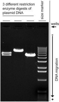
The more dense a band is, the more material there is.
With the gel that I ran, shown below, the left-most column was a mistake, and is to be disregarded. The second from the left is a negative control. It was basically only dye, to have a scale. The third is the positive control. The fourth and final column was my experiment. It was looking for a region called DMR. You see that band at the bottom of the the final one? That is DMR. I found it. However, the problem is the control. It is blurred and crazy. We have no idea what exactly went wrong there.

So the DMR is good, but the control is messed up. We can keep the DMR that I made in PCR, but have to run the control again, else we won't be able to actually experiment with it.
AB decided that he would set up the control for PCR so it would be quicker, and if I noticed anything that he did differently, then we might know what went wrong.
Start to finish, he was done in 15 minutes. He did half of what I did, but when I did it, it took me an 1.5 hours. This means that he did it three times faster than me. I guess practice makes perfect.
LATER: It is now four hours after I wrote that up. AB's new PCR is done. We also ran the gel electrophoresis. We took a picture. You know what? His looks just the same as mine.
This means a number of things. First and foremost, I didn't mess everything up. The other really big thing, is that we have no idea what is wrong. We have pretty much found that it wasn't really the mixing that messed things up. It might be either the quality or quantity of the reagents that went into it. Also, it might be something with the temperature of the thermocycler.
Next week, we will try to address this problem. Possibly, there was too much template DNA. This is odd because this PCR has been successful twice before, but has now failed twice. Possibly, it means that the amount of template DNA is right on the threshold- in the right conditions it might be enough but under the wrong conditions it's too much. We have no idea what the "right conditions" actually are.